|
|
|
|
|
|
| |
Molecular “mapping” uses powerful next-generation microscopes to create diagrams of cells, genes, and even entire organs to better understand how they work within the body and to develop targeted disease treatment. |
|
| |
|
|
|
|
|
|
|
| |
| |
|
|
| |
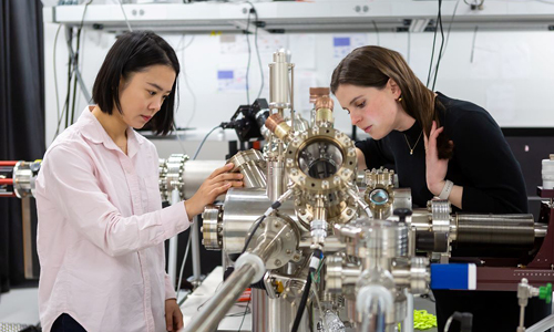
Assistant professor of chemistry Sarah King (right) and a student working with a photoemission electron microscope. (Photography by Nancy Wong) |
|
| |
|
|
|
|
| |
|
|
|
| |
Neurobiologists have long aimed to develop a detailed wiring diagram called the “connectome” that maps the connections among the brain’s 100 billion neurons. Researchers from three Chicago universities recently discovered a new way around one of the major challenges facing this effort: the slow imaging speeds of current microscopes. By adapting photoemission electron microscopy (PEEM), typically used in materials science, they are able to capture ultrathin brain slices more efficiently. Once processed, the data will be shared in online repositories, enabling experts and students to conduct further research and uncover new insights into brain anatomy and function. |
|
| |
|
|
|
|
|
| |
RNA, receptors, and randomness |
|
| |
|
|
|
| |
|
|
|
| A topical breakthrough: A new method of single-cell RNA sequencing, called TopicVelo, uses static data to understand how cells and genes change over time. |
|
|
|
| |
|
|
|
|
|
| |
|
|
|
| First look: Researchers have mapped the complete structure of a key protein involved in cell communication for the first time, revealing its adaptive function and opening possibilities for targeted drugs to treat diseases like cancer and brain disorders. |
|
|
|
| |
|
|
|
|
|
| |
|
|
|
| Bright-eyed and heavy-tailed: A study from UChicago, Harvard, and Yale found that neuronal networks across species exhibit a “heavy-tailed” distribution, suggesting shared principles of brain network organization. |
|
|
|
| |
|
|
|
|
|
| |
A blueprint to beat cancer |
|
| |
|
|
|
|
|
| |
| |
|
|
| |
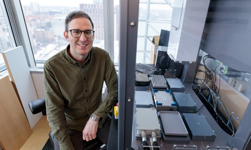
Associate professor of molecular engineering Nicolas Chevrier in his lab, which develops interdisciplinary approaches and tools to study how the immune system functions across biological scales. (Photography by John Zich) |
|
| |
|
|
|
|
| |
|
|
|
| |
A new platform for high-resolution spatial profiling of tumor microenvironments is poised to revolutionize cancer research. Developed in the lab of Nicolas Chevrier, associate professor at the Pritzker School of Molecular Engineering, the system uses microscope slides outfitted with a million tiny probes to analyze tissue samples as large as whole organs. Unlike previous profiling methods, the platform analyzes samples with higher sensitivity and resolution—but at a fraction of the cost. Chevrier seeks to use the platform to study archived biospecimens and large patient cohorts to uncover new biomarkers and therapeutic targets for cancer treatment. |
|
| |
|
|
|
|
|
|
|
| |
| |
|
|
| |
Trusting the gut:
The trillions of microbes in your gut not only aid digestion but also influence immunity, metabolism, allergies, and even neurological health.
|
| |
|
|
|
| |
New institute to combat climate change:
The University of Chicago launched the Institute for Climate and Sustainable Growth to take on the intertwined challenges of climate change and economic prosperity.
|
| |
|
|
|
|
|
|
| |
|
|
|
|
|
| |
| |
Sign up to receive µChicago monthly. |
|
|
|
| |
|
|
|
|